A model organism – the virtual bacteria.

stanfordmycoplasmagenitaliumI was reading an article in Scientific American today that got me thinking about the complexities of biology – the article described the production of a virtual bacteria using computing to model all of the known functions of a simple single cell.  The article was a very compelling read and presented a rational argument of how this could be achieved, based on modelling of a single bacterium that would eventually divide.  The benefits of a successful approach to achieving this situation are immense for both healthcare and drug development, but the difficulties are equally immense.

In order to simplify the problem the organism chosen to be modelled is a bacterium with the smallest genome size – Mycoplasma genitalium a bacteria that has only 500+ genes all of which have been sequenced.  This bacterium is also medically important, which adds weight to the usefulness of a virtual organism.  The problem of programming the computer was divided into modules, each of which will describe a key process of the cell all of which could feedback in a way that described actual binding coefficients for protein-protein and protein-DNA interactions.

As I read the article, I began to realise that there were some simple problems associated with the description of how the computer would “manage” the cell and when the author described doubling protein concentrations prior to cell division I knew there were problems with their model – this simplistic approach is not what happens in the cell – cellular control is an important aspect of this modelling and must be correct if the cell is to be realistic.  I can illustrate what I mean with one example – plasmid replication.  A plasmid in an autonomous circle of DNA, often found in bacteria, that are easy to understand and ubiquitous in nature.

Replication of plasmid DNA:

ReplicationThe number of copies of a plasmid are tightly controlled in a bacterial cell and this control is usually governed by an upper limit to an encoded RNA or protein, when the concentration of this product drops, such as AFTER division of the cell, replication will occur and the number of plasmids will increase until the correct plasmid number is attained (indicated by the concentration of the controlling gene product).

This is a classic example of cellular control and is a lot different to a model based on doubling protein concentration in anticipation of cellular division.

Mycoplasma genetics and the complexity of biology:

This whole problem also got me thinking about my own subject area and my brief excursion into the study of restriction enzymes from Mycoplasma. Restriction systems are a means for bacteria to protect themselves from viral invasion and, despite the small size of the genomes of Mycoplasma bacterial strains, they encode such systems.  There is clear evidence that such systems are “selfish” and may be they are fundamental to the long term survival of bacteria, so I think they need to be dealt with in the model organism.  However, things begin to get complicated when you look at the well described system from Mycoplasma pulmonis (a slightly more complex version of the organism used for the model).  Instead of a single set of genes for a restriction system, as usually found in other organism, the restriction genes of Mycoplasma pulmonis  are capable of switching in a way that can generate four different proteins from a single gene.  This is where the complexity of modelling an organism occurs and while the organism used may have a simple genome, it is important to know of how even simple organisms can increase their genetic complexity without increasing their DNA content.

Conclusion:

I think the work at Stanford is both interesting and important and I think they have achieved a very important step along the road to modelling a living cell, but I also think they may need more information and have more complex modules available to them as they try to be more accurate with even these simple organisms.  It will be a long road before we have a model of a human cell, but what an incredible thought that would be!


Synthetic Biology – will it work?

Eng_Future_Logo_OutlinesEvery now and then science comes up with a new approach to research that impacts on technology, but often these approaches are controversial and the headlines we see are far from the truth and can damage the investment into the new techniques.  One good example is the Genetic Modification of plants and the production of GM-foods, which has a really bad press in Europe despite many obvious benefits for the world economy and for disease control.  The latest technology, which follows from the explosion in genetic engineering techniques during the 1990s, builds on concepts developed in bionanotechnology and is known as Synthetic Biology.  But, what is Synthetic Biology?  Will it work?  And what are the dangers versus benefits of these developments?  Gardner and Hawkins (2013) have written a recent review about this subject, which made me think a blog on the subject was overdue.

My background in this area is two-fold:

  1. I was a part of a European Road-Mapping exercise, TESSY, that produced a description of what Synthetic Biology is and how it should be implemented/funded in Europe.
  2. I was also Project Coordinator for a European research project – BioNano Switch, funded by a scheme to support developments in Synthetic Biology, that aimed to produce a biosensor using an approach embedded in the concepts of Synthetic Biology.

So, what is Synthetic Biology?  I think the definition of this area of research needs to be clearly presented, something that was an important part of the TESSY project, as the term has become associated simply with the production of an artificial cell.  However, that is only one small aspect of the technology and the definition TESSY suggested is much broader:

Synthetic Biology aims to engineer and study biological systems that do not exist as such in nature, and use this approach for:

  • achieving better understanding of life processes,
  • generating and assembling functional modular components,
  • developing novel applications or processes.

syntheticBiologyThis is quite a wide definition and is best illustrated with a simple comparison – in electronic engineering there exists a blueprint (circuit diagram) that shows how components (resistors, capacitors etc.) can be fitted together in a guaranteed order to produce a guaranteed result (a device such as an amplifier).  The Synthetic Biology concept would be to have a collection of such components (DNA parts that include promoters, terminators, genes and control elements; cellular systems including artificial cells and genetically engineered bacteria capable of controlled gene expression; interfaces that can connect biological systems to the human interface for useful output).  This would mimic the electronic situation and provide a rapid mechanism for assembly of biological parts into useful devices in a reliable and predictable manner.  There are many examples of such concepts, but the best known is the Biobricks Foundadtion.  However, at the TESSY meeting I was keen to make it clear that there are fundamental problems with this concept, so what are the problems?

At its most simple concepts a Biobricks database would consists of a number of different types of DNA (promoters, are short DNA sequences that switch a gene on; terminators, are short DNA sequences that switch a gene off; control elements, are DNA sequences that control the promoter switching on or off a gene as required; genes, would be DNA sequences that produce Recombinant DNA
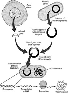
biotechnologically useful products; and cells, are the final package that enables the DNA to do its work and produce the required product), which sounds logical and quite simple.  However, biological systems are not as reliable as electronic systems and combinations of promoters and genes do not always work.  One of the major problems with protein production, using such artificial recombinant systems, is protein aggregation resulting in insoluble proteins that are non-functional.  In addition, there are many examples (usually unpublished) of combinations of Biobricks that do not work as expected, or if used in a different order also result in protein aggregation, none of which ever happens with electronic components.  The reasons are far from clear, but are closely related to the complexity of proteins and the need for them to operate in an aqueous environment.  My thoughts about how to deal with this situation is to have a large amount of metadata associated with any database of Biobricks, which includes information about failures or problems of protein production from specific combinations.  However, I am not aware of any such approach!

Synthetic CellThere are other aspects of Synthetic Biology that do not depend on Biobricks and one example is the artificial cell.  The ideal for such a system is a self-assembling package, capable of entrapping DNA, capable of replication and survival and able to produce useful biomaterials and significant steps have been made toward such a system.  However, one area of concern as such systems are developed, is containment – can we really be sure these artificial microbes will remain in a contained environment and not escape to interact with and possible change the natural bacterial population.  However, the power and capability of such a system should not be underestimated and the likely use in future medicine could be immense – simple examples would be as delivery systems for biomaterial that can activate cellular changes by targeting to the required cell and then switching on protein production (e.g. hormones).  This type of targeted medicine would be a major breakthrough during the later part of this century.

SEN25_BIO11Another type of Synthetic Biology involves the artificial assembly (possible self assembly) of biomaterials onto an artificial surface in an way that is unlikely to occur naturally, but provides a useful device – I see this as more like what a Biobricks project should be like – such a system is usually modular in nature and the bio-material would normally be produced using recombinant techniques.  The research project I mentioned earlier involved such a device and the outcome was a single molecule biosensor for detecting drug-target interactions at the limits of sensitivity.  The major issues we had with developing this device was the precise and accurate attachment of biomaterial, to a surface in such a way that they function normally.  However, overall the project was successful and shows that a Synthetic Biology approach has merits.

What are the benefits that Synthetic Biology can provide society?  Well, one advantage is a more systematic approach to biotechnology, which to date has tended to move forward at the whim of researchers in Academia or industry.  Assuming the problems, associated with protein production, mentioned above can be better understood then there could be a major boost in use of proteins for biotechnology.  In addition, Synthetic Biology techniques offer a unique opportunity for miniaturisation and mass production of biosensors that could massively improve medical diagnosis.  Finally, artificial cells have many future applications in medicine, if they can be produced in a reliable way and made to work as expected:

  1. They could provide insulin for diabetics.
  2. Be made to generate stem cell, which could be used in diseases such as Alzheimer’s and Huntingdon’s.
  3. They could deliver specific proteins, drugs and hormones to target locations.
  4. They could treat diseases that result from faulty enzyme production (e.g. Phenylketonuria).
  5. They could even be used to remove cholesterol from the blood stream.

However, there are always drawbacks and risks associated with any new scientific advance:switch%20off

  1. Containment of any artificial organism is the most obvious, but this enhanced by the possibility of using the organism to produce toxins that would allow its use as a biological weapon.
  2. The ability to follow a simple “circuit diagram” for protein production, combined with a readily available database of biological material, could enable a terrorist to design a lethal and unpredictable weapon much more complex and perhaps targeted than anything known to date.
  3. Inhibit research through a readily available collection of materials that prevent patent protection of inventions.  This could be complicated by the infringement of patents by foreign powers in a way that blocks conventional research investment.
  4. Problems associated with the introduction of novel nano-sized materials into the human body, including artificial cells, which may be toxic in the long term.

My own feeling is that we must provide rigorous containment and controls (many of which already exist), but allow Synthetic Biology to develop,  Perhaps there should be a review of the situation by the end of this decade, but I hope that the risks do not materialise and that society can benefit from this work.

Prions, protein aggregation and disease development.

PrionImgPrions are believed to be the causative agents of a number of diseases that develop through protein aggregation (amyloidosis) including BSE (bovine spongiform encephalopathy) in cattle and CJD (Creutzfeldt-Jakob disease) in humans.  The formation of protein aggregates produces plaques within the normal neural cell structure, disrupting the cellular structure and resulting in a soft, spongy like appearance.  The result of these effects are varied and include dementia, convulsions, balance and coordination dysfunction and, in humans, disturbing personality changes.  Prions differ between species and, in general, cross species transfer is not infective of the disease.  However, CJD prion is thought to have been derived from the BSE prion, transmitted through contaminated meat.  Whether prions are the agent which causes the diseases, or are a symptom caused by a different agent, is still debated by some researchers.  However, the general consensus is that CJD and BSE are prion-based diseases.

prion_ilus340x312Prions are proteins that have two different states into which they can fold and it is this change in the folded state that leads to aggregation and development of the associated diseases.  Prions are found in many very different species and a recent study (Kim et al., Nature, 2013. 495(7442): p. 467-473) has identified key amino acids, conserved across prion proteins, which appear to be important in the aggregation process.  This suggests that a more focussed drug development project could be initiated, in which drug docking could be used to design drugs to interfere with the aggregation process.

Although such developments may still be a long way away, this is an exciting time where modern technologies could be used together to provide more breakthroughs in halting these diseases – I am for ever optimistic!

Thoughts on single molecule science

Some while ago I worked with two groups who were using magnetic tweezers systems to study single molecule biophysics and I was always keen to expand that work to new projects F1_medium(unfortunately retirement moved such thoughts away from actuality).  A recent paper, using single molecule FRET analysis (Haller, A., et al. Proceedings of the National Academy of Sciences, 2013. 110(11): p. 4188-4193) has investigated the folding of an RNA aptamer.

This work encourages me to suggest further studies of aptamer folding using magnetic tweezer systems with a long-term view of developing a sensor for aptamer activity – it would be incredible to develop a toxin sensor, based on aptamers to specific toxins, that detects at the single molecule level!

Fig 10This is not such an over-ambitious project and relates closely to my own work, with a large European team, to develop a single molecule biosensor.  The basis of the three European projects, involved in this work, was to develop the magnetic tweezer device as an automatic electronic platform for detection of magnetic bead movement based on changes to the length of DNA molecules located within the device.  The concept was to use the device to identify drug-target interactions involving DNA-manipulating enzymes (such as helicases and topoisomerases) that are potential targets in anti-cancer drug development.

However, I always envisioned a greater potential for such a device and studying aptamers would be only one aspect of such a project.  The device could also be used to study Quadruplex formation, and drugs that affect this, other fibre formation, such as amyloids and even protein-protein interactions involving DNA binding enzymes to enable DNA-loop formation as an indicator of such interactions.  All of these uses provide a potential health-orientated development of the biosensor – just need someone to have the enthusiasm to write the proposal!

Memories of scientific discussions – Raman Spectroscopy

Three Bulls HeadsMany years ago, on many a Friday lunchtime, I used to sit in The Three Bulls Heads, Newcastle discussing applications of Physics within Biology with a Physicist who also worked at The University (if he reads this he will know who he is!).  Those chats were often quite complex, covering lots of difficult chemistry, physics and biology – goodness knows what the regulars thought of us!

One subject area that I was very interested in was the possible applications of Raman Spectroscopy within biology and that subject has interested me for many years; although, I have never had much opportunity to apply the technology to any of my research, which is a shame.  So, the question arises, for my blog readers, what is Raman Spectroscopy and has it found uses in biology?

Interestingly, the whole concept of Raman Spectroscopy relates to what has become known as the wave-particle duality, which was one of my favourite tutorial subject that I brought to my unsuspecting biology undergraduates.  It is really difficult to visualise how a particle can be a wave and vice-versa, but I did my best to try to explain this and this was the basis of my explanation…..

250px-Wave_particle_duality_p_known_svgIf you think of a wave as a string vibrating very quickly then it is easy to see that where the movement of the string is maximum then this region could be “seen” as a particle.  If the vibration frequency is changed several waves form and this can be “seen” as several particles.  This is a little simplistic, but is also interesting as quantum mechanics shows that particles such as electrons and atoms can be described by a wave equation (Schrödinger).  The major different is that we have to imagine three dimensions, which is very difficult, but if you could then you would understand how particles exits as waves – I am not sure how well this explanation works, but it is the best I have come up with.

Raman-ScatteringSo where does Raman scattering fit in to this?  Well, Raman described the possibility that photons, as particles, could impact each other, or with other particles such as electrons, and be scattered in an inelastic manner ( a sort of back scattering).  The interaction of the incident light with electrons used to bond different atoms in a molecule results in scattered light of different wavelengths (Raman Spectroscopy is the measurement of this light).

scanner-30A recent paper (Brauchle & Schenke-Layland. Biotechnology Journal, 2013. 8(3): p. 288-297) has reviewed the current applications of Raman Spectroscopy within biology and suggested future applications.  This has brought me up to date following these discussions so many years ago and I am pleased eo hear that some of the ideas we had are now feasible, through improved instrumentation. Specific signals can be measured from proteins, lipids, carbohydrates, DNA etc., which makes it possible to show changes and abnormalities in these components  and detection of infections or disease.  It has already been demonstrated that this non-invasive technology can be used to identify bacterial infection, tumour development and cell death.  Undoubtedly, this technology will improve and maybe one day all of those Star Trek fans will see a body scanner that is predictive about well-being – who knows, lol!

Huntingdon’s disease – new thoughts and observation.

Although Huntingdon’s Disease (HD) is far from my area of expertise in science, I feel I have some background knowledge and, consequently, some interest after reading and commenting on many a treatise about this subject, for a an old colleague when I was working.

huntingtonsHD is a neurological disorder that eventually (usually in mid to late life) leads to cerebral atrophy (as illustrated), which manifests itself through involuntary movement, loss of cognitive  ability and psychiatric problems.  The genetic cause of the disease is the presence of CAG repeat sequences within the DNA of the HTT gene (within exon 1 of the gene).  These repeat triplets of DNA sequence are translated within the huntingtin protein (HTT) to produce a region of poly-glutamine, a highly charged region within the protein that disrupts normal function.  The timing of development of the disease is linked to the number of CAG repeats within the HTT gene, above 40 leads to normal (late) development of the disease, but more than 70 CAG repeats leads to disease development in childhood.HTT gene

The presence of the CAG repeats points to the N-terminal region of the HTT protein being the toxic species and small fragments of this part of the HTT protein have been found in analysis of patient’s brain tissue post-mortem.  These protein species appear to be small fragments released from the HTT protein and suggest proteolysis of the intact protein may produce the toxic species.  However, this concept was challenged in a recent paper by Sathasivam et al. (Sathasivam, Proceedings of the National Academy of Sciences 110(6): 2366-2370).

alternative_splicingOne possible alternative, which might produce short fragments of HTT protein, would be an alternative splicing mechanism that could generate short mRNA fragments that produce equally short protein fragments.  Splicing is a mechanism by which the intermediate between DNA (genes) and proteins – mRNA – is cut into fragments that define specific domains or regions of the final protein.  Alternative sites for the splicing mechanism can produce alterative proteins, or even protein fragments.  Sathasivam et al., have shown that splicing of exon 1 of the HTT mRNA occurs in a CAG repeat-length dependent manner, producing aberrant mRNA fragments that are translated into the short protein fragments found associated with diseased brain cells.

This is an important breakthrough in understanding the molecular basis for this disease, but also suggests that drugs targeted at reducing levels of HTT mRNA production (transcription of the gene) would not necessarily work as this would not necessarily reduce the amount of exon 1 mRNA available for aberrant splicing.  Therefore, a new direction for drug development may be required.

Could phage become useful viruses?

I remember many years ago my old professor telling me how bacteriophage could be useful tools for typing bacteria, based on their susceptibility to different restriction enzymes, which occur in different numbers in different bacteria, but, sadly, it never came to pass.  However, I read a recent paper (Henry & Debarbieux, 2012. Volume 434, 151–161) that suggest a new set of tools could be made available for healthcare that could make use of viruses and in particular bacteriophage.

bacteriophageSo, what are bacteriophage?  Well these are viruses that specifically infect bacteria and there thousands of them for all of the different bacteria known (and probably many more as yet unknown) and often when they do infect and kill the host, they use the bacterial replication system to copy themselves many times.  As rather beautifully illustrated on the left, they attach to the surface of a bacteria, using a highly specific attachment mechanism, and then inject their nucleic acid (DNA or RNA) into the cell like a biological hypodermic needle.  If grown artificially, in a laboratory, on a surface coated with growing bacteria, this produces areas of killing know as plaques – an example is illustrated at http://www.typei-rm.info/background.htm – the idea of typing bacteria is based on changes to the number of plaques by the defence systems bacteria use to stop phage growth – restriction enzymes.  Each strain of bacteria carries a different array of restriction enzymes, which should produce different efficiencies of growth of saturating concentrations of phage, sadly, the restriction enzymes are so efficient at killing the phage that growth show little variation between the many strains!

Since my early days in molecular biology, bacteriophage have been manipulated and the genomes have been deleted or changed to make use of them as the tools of molecular biology.  In fact, it is really the success of work by Prof Noreen Murray that initiated the ideas of using viruses as molecular tools; she was able to produce altered bacteriophage genomes that now carried “foreign” DNA and this produce some of the first genetically engineered organisms.

CaptureViruses are already used to deliver both drugs and to engineer genomes against disease, but how this might work and how useful viral tools might be best illustrated by a mechanism know as Phage Display, which has been used to produce artificial antibodies without recourse to injection of mice or other mammals!  The antibody proteins are attached to the outside of the bacteriophage, where they are functional, and they can be used to bind to an antigen attached to a surface (for example an antigen marker that is known to occur in cancer cells), the phage that bind can be isolated, manipulated genetically to improve binding to the antigen and then purified under more stringent procedures – panning.  Eventually, an antibody is isolated that is a tight binder to the antigen and could be used therapeutically.  However, one can imagine the next step in this type of work would be to target viruses at cancer cells, using viral display of specific antibodies, and then kill the cancer cells using part of the viruses normal life cycle.

So, do viruses hold a useful future for genetic engineers and bionanotechnologists?  I would say, as we begin to fully understand the genetics of the many viruses and learn to manipulate the genomes in a safe way, we could very well have a series of tools for manipulating genetic information, or for that detection, diagnoses, control and even cure of infectious diseases.  Who knows whether in another decade, during which time many new viruses will be discovered, the possibilities are enormous!

The role of RNA in Biology

The Central Dogma of Molecular Biology, DNA is transcribed into mRNA, which is translated into a protein.

If you are a biologist, and especially a molecular biologist, then you know all about the Central Dogma and the role of RNA as the intermediate between DNA (coding for genes) and proteins (the final product of most genes).

There are a number of species of RNA, including messenger RNA (mRNA) that is the product of transcription and is produced by RNA Polymerase (RNAP), while transfer RNA (tRNA) is used by the ribosome during translation as the mechanism for decoding the RNA sequence into a protein sequence and ribosomal RNA (rRNA) is a major constituent of the ribosome providing both structural form and enzymatic activity required for protein synthesis.
However, there are other types of RNA and perhaps one type that has become very well-known in recent years is small interfering RNA (siRNA).  These short RNA sequences were first discovered (at the end of the 20th Century -Hamilton & Baulcombe (1999). Science 286, 950-952) as molecules capable of silencing gene expression in plants.  Closely related to siRNA are microRNA (miRNA), these are also short (20+ nucleotides long) and occur in many copies within a genome, but are capable of regulating a large number of genes through a system involving the guidance of Argonaute proteins to silence specific genes.  This led to them also being known as silencing RNAs and to a massive expansion of interest in small RNA molecules. Their potential for therapeutic and biotechnological uses is summarised in a recent paper (Glorioso, J.C. (2011).  Gene Therapy (2011) 18, 1103).
The mechanism of action is that the small RNA molecules, which are released from a pre-miRNA transcript of specific genes through a series of processing steps involving an exoribonuclease and export to the cytoplasm, bind specific pockets or folds within the Argonaute proteins and lead this protein to attach to specific mRNA and silence them, or mark them for destruction.  Silencing is through binding of the miRNA-Argonaute protein complex to the 5′-untranslated region (UTR) of the mRNA and only 6-8 complementary bases are required between MiRNA and mRNA for silencing to occur.  mRNA destruction occurs by the Ago2, which is closely related to RNse H and the only Argonaute protein that retains this nuclease activity, but requires complete complementarity between the miRNA and the UTR region of the mRNA.  Coding regions of the genome represent only a very small part (a few percent) of the total genome size and it now clear that interference RNA is one example of a large number of RNA molecules preesent in the non-protein-coding regions of the genome (small nucleolar RNAs (snoRNAs), PIWI-interacting RNAs (piRNAs), large intergenic non-coding RNAs (lincRNAs) to name just a few).  Esteller, (2011) has detailed the close link between these non-coding RNA molecules and  human disease (Nat Rev Genet 12: 861-874), but it is clear that much work is required to identify all of these non-coding RNA molecules and identify their targets and how this relates to disease.  
miRNA production is closely linked to specific cell types and various stages of cellular differentiation, while unexpected production of miRNA molecules is linked to a number of disease states including cancer, diabetes, hearing loss and liver disease.  Certain viruses (particularly from the Herpes virus family) produce a number of miRNA molecules , which they use to overcome the human immune system by targeting the major Histocompatibility complex class I chain-related molecule B (a natural killer cell ligand).  Interference with these miRNA molecules could enhance natural immunity to herpes viruses.  Both increasing the levels of miRNA in the cell and introduction of miRNA molecules, or their complements, has been found to influence the development of diseases and the development of cancer.  miRNA and siRNA molecules, that bind targets that are difficult to treat with normal drugs (such transcription factors like MYC), may represent a way to inhibit new targets for control of cancer and an exciting way forward for disease treatments in the future.
A key aspect of using interference RNA for disease treatment will be targetted delivery of a source of these nucleic acid molecules and a variety of methods for transfecting cells have been explored (Davidson & McCray (2011) Nat Rev Genet 12: 329-340).  These include using bacteria to deliver specially designed DNA molecules as a source of siRNA, lipid encapsulation of the molecules and viral delivery systems.  Mutation and subsequent resistance to the treatment is a known problem as is natural degeneration of these moleules and a consequent need for repaeated treatment.  Yet this area has moved a long way in only a decade and is a serious area for future therapies.

The role of RNA in Biology

The Central Dogma of Molecular Biology, DNA is transcribed into mRNA, which is translated into a protein.

If you are a biologist, and especially a molecular biologist, then you know all about the Central Dogma and the role of RNA as the intermediate between DNA (coding for genes) and proteins (the final product of most genes).

There are a number of species of RNA, with messenger RNA (mRNA) being the product of transcription and is produced by RNA Polymerase (RNAP), while transfer RNA (tRNA) is used by the ribosome during translation as the mechanism for decoding the RNA sequence into a protein sequence and ribosomal RNA (rRNA) is a major constituent of the ribosome providing both structural form and enzymatic activity required for protein synthesis.
However, there are other types of RNA and perhaps one type that has become very well-known in recent years is small interfering RNA (siRNA). These short RNA sequences were first discovered (at the end of the 2oth Century -Hamilton & Baulcombe (1999). Science 286, 950-952) as molecules capable of silencing gene expression in plants. Closely related to SiRNA are MicroRNA (miRNA), these are also short (20+ nucleotides long) and occur in many copies within a genome, but are capable of regulating a large number of genes through a system involving the guidance of Argonaute proteins to silence specific genes. This led to them also being known as silencing RNAs and led to a massive expansion in interest in small RNA molecules and their potential for therapeutic and biotechnological uses is summarised in a recent paper (Glorioso, J.C. (2011). Gene Therapy (2011) 18, 1103).
The mechanism of action is that the small RNA molecules, which are released from a preMiRNA transcript of specific genes through a series of processing steps involving an exoribonuclease and export to the cytoplasm, bind specific pockets or folds within the Argonaute proteins and lead this protein to attach to specific mRNA and silence them, or mark them for destruction. Silencing is through binding of the MiRNA-Argonaute protein complex to the 5′-untranslated region (UTR) of the mRNA and only 6-8 complementary bases are required between MiRNA and mRNA for silencing to occur. mRNA destruction occurs by the Ago2, which is closely related to RNase H and the only Argonaute protein that retains this nuclease activity, but requires complete complementarity between the MiRNA and the UTR region of the mRNA.
MiRNA production is closely linked to specific cell types and various stages of cellular differentiation, while unexpected production of MiRNA molecules is linked to a number of disease states including cancer, diabetes, hearing loss and liver disease. Certain viruses (particularly from the Herpes virus family) produce a number of MiRNA molecules , which they use to overcome the human immune system by targeting the major Histocompatibility complex class I chain related molecule B (a natural killer cell ligand). Interference with these MiRNA molecules could enhance natural immunity to herpes viruses. Both increasing the levels of MiRNA in the cell and introduction of MiRNA molecules or their complements has been found to influence the development of diseases and the development of tumours (Broderick & Zamore (2011).  Gene Therapy 11, 1104-1110).  Clinical trials of MiRNA-complementary short oligonucleotides has already begun and this area promises a strong future research area.
I imagine over the next few years that the study of how short RNA molecules control gene expression and how they may effect disease development will show massive progress with a huge potential for new treatments (Kasinski & Slack (2011).  Nature Reviews Cancer 11, 849-864).